The heart is a muscle whose job is to pump blood around the body. The heart is the main organ of the circulatory system. The circulatory system also contains a network of arteries, veins and capillaries which deliver blood to different parts of the body.
Blood carries oxygen and nutrients to your organs and carries carbon dioxide to the lungs to be exhaled. The heart is powered by electrical impulses sent by the brain and nervous system. The impulses make each chamber of the heart contract to squeeze blood from one area to the next, eventually pumping blood out of the heart to the rest of your body.
A healthy heart supplies your body and organs with blood at the right heart rate to help it work well. If your heart is not functioning properly, the body’s organs may not receive enough blood to work well.
Anatomy of the heart
The heart is located in your chest and is protected by your ribs and breastbone (sternum). It is slightly off-centre, on the left side of the body. It is about the size of your fist.
Your heart is divided into four sections or chambers. The two chambers on the top are called atria and the two chambers on the bottom are called ventricles. A muscular wall called the septum separates the right side of the heart from the left.

On the right side of the heart, there is a right atrium and a right ventricle. The right side receives oxygen-poor blood returned from the rest of the body.
On the left side of the heart, there is a left atrium and a left ventricle. The left side receives oxygen-rich blood from the lungs.
The four chambers are separated by one-way valves that open and close with every heartbeat.
There are four heart valves:
- Aortic valve
- Tricuspid valve
- Pulmonary valve
- Mitral valve
These one-way valves keep the blood flowing in one direction through the different chambers of the heart and out to the body. The heartbeat that can be heard with a stethoscope is the sound of your valves opening and closing to let blood through.
Valves that don’t work properly can lead to different types of valvular heart disease.
How does the heart work?
To pump blood throughout the body, your heart contracts then relaxes. This action is similar to clenching and unclenching your fist. With each time your heart contracts (beats), blood is being pushed through your arteries. This pumping in the large arteries of your body feels like a pulsing sensation which is what you feel when you take your pulse.
When the heart beats, blood flows in one direction. This is because of a system of valves that work to control the direction of flow. These valves open and close with each heartbeat.
Arteries deliver oxygen-rich blood to all parts of the body and veins bring the deoxygenated blood back to the lungs. Small vessels called capillaries run from the arteries and veins into the organs.
Like any other muscle, the heart needs its own supply of oxygen-rich blood to function. The coronary arteries handle this job. Coronary artery disease is when the coronary arteries become narrowed or blocked so your heart does not get enough oxygen to do its job effectively. When the heart is impaired, it struggles to provide blood to the body’s organs.
Heart disease refers to four groups of heart conditions that can affect the structure and functions of the heart.
- coronary artery and vascular disease
- heart rhythm disorders (arrhythmias)
- structural heart disease (congenital heart disease, heart valve disease)
- heart failure

How blood flows through the heart
When the heart beats, blood follows a predictable, one-way path through the heart and circulatory system.
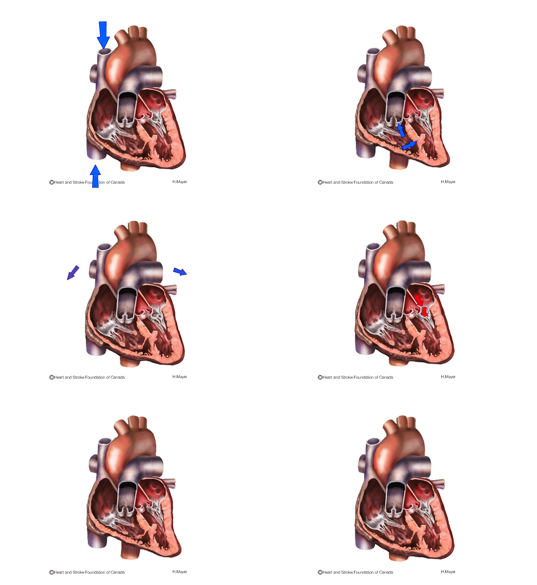
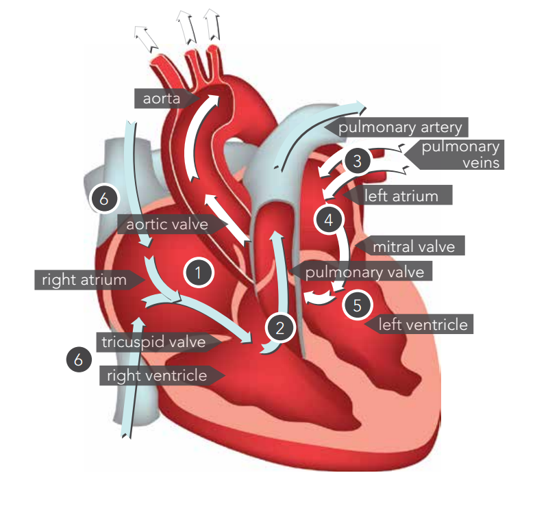
- The right atrium is full of oxygen-poor blood returned from your body (muscles, organs, brain and heart). When the right atrium becomes full, it contracts. When the atrium contracts, the tricuspid valve between the right atrium and the right ventricle opens. The blood flows into the right ventricle.
- When the right ventricle is full it contracts and pumps the blood to the lungs through the pulmonary valve.
- In the lungs, carbon dioxide is removed and fresh oxygen is added to the blood. The blood is now oxygen-rich. Oxygen-rich blood then flows into the left atrium.
- When the left atrium contracts, the mitral valve between the left atrium and left ventricle opens. The blood flows into the left ventricle.
- The left ventricle pumps the oxygen-rich blood into the aorta through the aortic valve and out to the rest of your body.
- The oxygen-rich blood travels throughout arteries in your body. Veins carry the oxygen-poor blood back to the right atrium. The right atrium fills with oxygen-poor blood, and then the cycle begins again.
What is a healthy heart rate?
Your heart rate is the number of times your heart beats per minute. Normal heart rate varies from person to person, but a typical adult resting heart rate is usually about 60 to 100 beats per minute.
During rest, your heartbeat will slow down. This measurement is known as your resting heart rate. With exercise, the heart will beat faster. Being aware of your heart rate can help you spot health problems. Many smartwatches and fitness trackers can measure your heart rate throughout the day.
Both the rate (speed) and rhythm of your heart are important for healthy heart function. A normal heart rhythm is called normal sinus rhythm (NSR). Heart beats that are too slow (bradycardia, less than 60 beats per minute) or too fast (tachycardia, more than 100 beats per minute) are two types of arrhythmias.
How does the heart conduct electricity?
The rhythm and pace of the heartbeat is controlled by an electrical conduction system which transfers signals from the brain and nervous system to the heart. This system helps make sure the heart beats at a normal rate and in a regular rhythm.
The heart’s electrical system consists of nodes, branches and fibres, all of which work together to help the heart to beat.
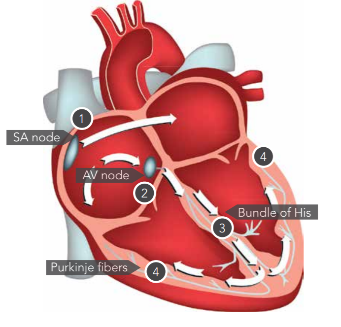
The SA node (sinoatrial node) is a structure that stimulates contraction of the heart. This initial electrical signal spreads throughout the heart from top to bottom (from atria to ventricles). As one part contracts, the others relax.
The electrical branches and fibres participate in the process, sending electrical impulses to different parts of the heart.

The heart’s electrical system
- The electrical signal travels from the SA node to the atria causing them to contract pumping blood into the ventricles.
- The signal then goes to the AV node (atrioventricular node) between the atria and the ventricles. The signal is held there for a moment while the blood is being pumped into the ventricles.
- Then the signal moves to the ventricles through fibres inside the septum and ventricle walls called “bundle of His” and “Purkinje fibres”.
- This makes the ventricles contract – sending blood to the lungs and to the rest of your body.
An ECG or EKG (electrocardiogram) is a test that checks how your heart is functioning by measuring the electrical activity that passes through the heart. A trained health care provider can determine if this electrical activity is normal or irregular.
How can I keep my heart healthy?
There are many different conditions that can affect the heart. However, there are many ways you can help keep your heart healthy through lifestyle choices and other behaviours:
- be smoke-free
- be more active
- eat a healthy balanced diet – there are some specific diets you can follow that have been proven to reduce the risk of heart disease
- maintain a healthy weight
- drink less alcohol
- avoid recreational drug use
- manage stress.
Talk to your healthcare provider about the lifestyle changes that will benefit you the most.
